
Page 450 - Ultrassonografia Aplicada à Dermatologia e à Cosmiatria - Uma Abordagem Clinica e Ecográfica
P. 450
ULTRASSONOGRAFIA APLICADA À DERMATOLOGIA E À COSMIATRIA
1
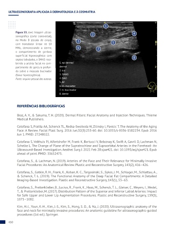
Figura 53. Jowl. Imagem ultras- 4
sonográfica (corte transversal), 2
no Modo B (escala de cinza), 3
com transdutor linear de 18 5
MHz, demonstrando a derme, 6 7
o compartimento de gordura
superficial hipoecogênico com
septos lobulados, o SMAS reco- 8
brindo a artéria facial no com- 1. epiderme/
partimento de gordura profun- derme
do sobre o músculo bucinador 2. tcs
(faixa hipoecogênica). 3. SMAS
Fonte: arquivo pessoal das autoras. 4. DAO
5. AF
6. m. masseter
7. m. bucinador
8. dente
REFERÊNCIAS BIBLIOGRÁFICAS
Braz, A. V., & Sakuma, T. H. (2020). Dermal Fillers: Facial Anatomy and Injection Techniques. Thieme
Medical Publishers.
Cotofana S, Fratila AA, Schenck TL, Redka-Swoboda W, Zilinsky I, Pavicic T. The Anatomy of the Aging
Face: A Review. Facial Plast Surg. 2016 Jun;32(3):253-60. doi: 10.1055/s-0036-1582234. Epub 2016
Jun 1. PMID: 27248022.
Cotofana S, Velthuis PJ, Alfertshofer M, Frank K, Bertucci V, Beleznay K, Swift A, Gavril D, Lachman N,
Schelke L. The Change of Plane of the Supratrochlear and Supraorbital Arteries in the Forehead - An
Ultrasound-Based Investigation. Aesthet Surg J. 2021 Feb 28:sjaa421. doi: 10.1093/asj/sjaa421. Epub
ahead of print. PMID: 33652475.
Cotofana, S., & Lachman, N. (2019). Arteries of the Face and Their Relevance for Minimally Invasive
Facial Procedures: An Anatomical Review. Plastic and Reconstructive Surgery, 143(2), 416–426.
Cotofana, S., Gotkin, R. H., Frank, K., Koban, K. C., Targosinski, S., Sykes, J. M., Schlager, M., Schlattau, A.,
& Schenck, T. L. (2019). The Functional Anatomy of the Deep Facial Fat Compartments: A Detailed
Imaging-Based Investigation. Plastic and Reconstructive Surgery, 143(1), 53–63.
Cotofana, S., Pretterklieber, B., Lucius, R., Frank, K., Haas, M., Schenck, T. L., Gleiser, C., Weyers, I., Wedel,
T., & Pretterklieber, M. (2017). Distribution Pattern of the Superior and Inferior Labial Arteries: Impact
for Safe Upper and Lower Lip Augmentation Procedures. Plastic and Reconstructive Surgery, 139(5),
1075–1082.
Kim, H.-J., Youn, K.-H., Kim, J.-S., Kim, S., Hong, S. O., & Na, J. (2020). Ultrasonographic anatomy of the
face and neck for minimally invasive procedures: An anatomic guideline for ultrasonographic-guided
procedures (1st ed.). Springer.
450